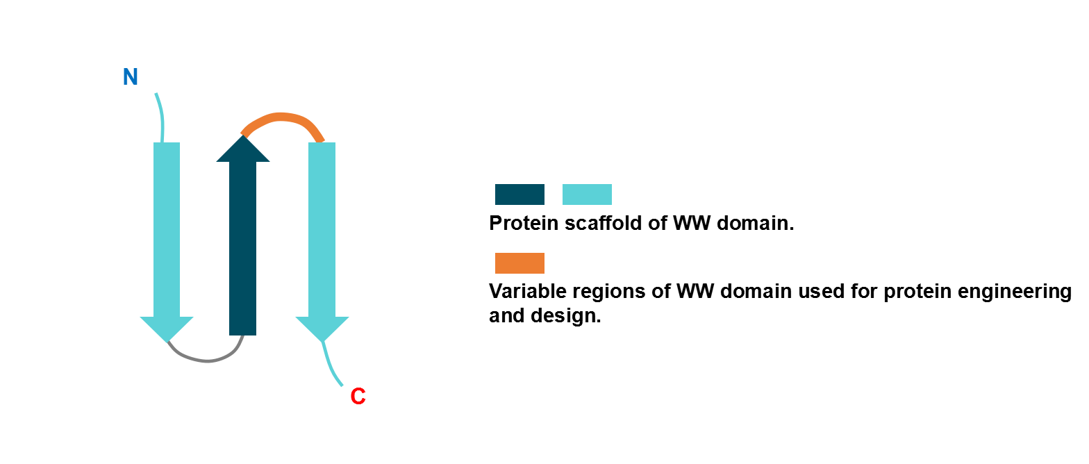
General Information of Protein Scaffold of SBP (PSS) (ID: PS067)
PSS Name
WW domain
Synonyms
WW domain-based binder
Diagrammatic Sketch
Highest Status Research
Class Non-Antibody
Fold Type Beta-Sheets + Loops
Thermal Denaturation TEMP 50-70℃
Sequence Length 36-57
Molecular Weight 4-8 kDa
Parental Protein of This PSS
Parental Protein Name Synonym PDB ID UniProt ID Origin
WW domain of Yes-associated protein WW domain of hYAP
2IDH
P46938 ; P46937
Human; Mouse; Synthetic construct
Synthetic Binding Protein (SBP) Featuring this PSS
SBP Name Highest Status Template Expression System Selection Method Details Ref
WW domain anti-peptide mutant PK1 Research hYAP65 Escherichia coli BL21 (DE3) Phage display
SBP Info
[1]
WW domain anti-peptide mutant PK2 Research hYAP65 Escherichia coli BL21 (DE3) Phage display
SBP Info
[1]
WW domain anti-peptide mutant PK3 Research hYAP65 Escherichia coli BL21 (DE3) Phage display
SBP Info
[1]
WW domain anti-peptide mutant PK4 Research hYAP65 Escherichia coli BL21 (DE3) Phage display
SBP Info
[1]
WW domain anti-Zn(II) WW-CA Research N.A. N.A. N.A.
SBP Info
[2]
WW domain anti-peptide mutant W17F Research hYAP Escherichia coli BL21 (DE3) N.A.
SBP Info
[3]
WW domain anti-VEGFR-2 clone B1 Research Peptide N.A. CIS display
SBP Info
[4]
WW domain anti-VEGFR-2 cycB1_4-28 Research Peptide N.A. CIS display
SBP Info
[4]
WW domain anti-VEGFR-2 cycB1_6-36 Research Peptide N.A. CIS display
SBP Info
[4]
WW domain anti-peptide mutant L30K Research hYAP65 N.A. N.A.
SBP Info
[5]
References
1 Evolution of binding affinity in a WW domain probed by phage display. Protein Sci. 2000 Dec;9(12):2366-76.
2 Switchable Zinc(II)-Responsive Globular -Sheet Peptide. ACS Synth Biol. 2022 Jan 21;11(1):254-264.
3 Characterization of the structure and function of W --> F WW domain variants: identification of a natively unfolded protein that folds upon ligand binding. Biochemistry. 1999 Oct 26;38(43):14338-51.
4 Selection of a high-affinity WW domain against the extracellular region of VEGF receptor isoform-2 from a combinatorial library using CIS display. Protein Eng Des Sel. 2013 Apr;26(4):307-15.
5 Solution structures of the YAP65 WW domain and the variant L30 K in complex with the peptides GTPPPPYTVG, N-(n-octyl)-GPPPY and PLPPY and the application of peptide libraries reveal a minimal binding epitope. J Mol Biol. 2001 Dec 14;314(5):1147-56.